Coordinator
Dr K.G.A. Gilhuijs (Kenneth)
Contact
Dr K.G.A. Gilhuijs (Kenneth) (K.G.A.Gilhuijs@umcutrecht.nl)
Faculty
Dr Kenneth Gilhuijs
Dr Alexander Leemans
Date
September – January; for the schedule please check the website of the Master’s programme Medical Imaging
Register by sending an e-mail to ImagO.
Please note: this course may be partly taught online in MS Teams. To access the Teams account you need a solis id. If you do not yet have one, apply for one on time.
Location
University Medical Center Utrecht
Course content
This course covers the full roadmap from basic to more advanced techniques that are commonly used in medical image procesing.You will learn how to analyse concrete medical questions that arise from medical images, and how they can be solved by mathematical analysis of CT, MRI and X-ray. We will take you from theory to design of computer-aided diagnosis systems and Radiomics systems. Examples of such systems are those that automatically detect tumors in CT and MRI scans, that automatically detect micro-aneurysms in retinal images, or that estimate the prognosis of breast-cancer patients based on imaging features that cannot be picked up by the human eye.
Topics include segmentation (dynamic programming, active contours, level sets), image registration, mathematical morpholpgy, texture analysis, pattern recognition (feature spaces, classifiers, support-vector machines and random forests). During the lectures we will provide small practical assignments using a voting system. A computer practicum (using matlab) will be provided to get hands-on experience with the different techniques. In addition, individual assignments are provided consisting of actual problems that were encountered in medical images.
Upon completion of the course the participant
- is able to choose the most appropriate technique for medical image processing and image analysis
- knows the underlying theory to understand the strenghts and weaknesses of common techniques for image segmentation, image registration, image feature extraction and image feature classification
- is able to evaluate image processing and analysis technniques using standardized methodology
- is able to implement solutions for new medical imaging problems
- knows the benefits and pitfalls of computer-aided diagnosis
Credits
You will be awarded 3 EC for attendance and completing the assignments (written report and presentation); a further 2 EC can be earned on passing the final exam.
Literature
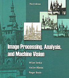
“Image Processing, Analysis and Machine Vision”, (Third edition) Author: M. Sonka
ISBN: 049508252X hard cover; ISBN: 0495244384 paperback
Highly recommended:
“Essential C++”, Author: S.B. Lippman, ISBN 0201485184
